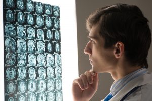
X-rays- a type of radiation called electromagnetic waves. X-ray imaging creates pictures of the inside of your body. The images show the parts of your body in different shades of black and white. Calcium in bones absorbs x-rays the most, so bones look white. The most familiar use of x-rays is checking for fractures (broken bones), but x-rays are also used in other ways; for example, chest x-rays can spot pneumonia. Mammograms use x-rays to look for breast cancer.
MRI’s- also known as magnetic resonance imaging. A medical imaging technique that uses a magnetic field and computer-generated radio waves to create detailed images of the organs and tissues in your body. A non-invasive way for your doctor to examine your organs, tissues, and skeletal system. MRI is the most frequently used imaging test of the brain and spinal cord. It’s often performed to help diagnose:
- Aneurysms of cerebral vessels
- Disorders of the eye and inner ear
- Multiple sclerosis
- Spinal cord disorders
- Stroke
- Tumors
- Brain injury from trauma
CT scan with and without contrast- also known as computerized tomography. A CT scanner emits a series of narrow beams through the human body as it moves through an arc. It can see tissues within a solid organ. This data is transmitted to a computer, which builds up a 3-D cross-sectional picture of the part of the body and displays it on the screen. Sometimes, a contrast dye is used because it can help show specific structures more clearly.
PET scan- also known as positron emission tomography. An imaging test that can help reveal the metabolic or biochemical function of your tissues and organs. The PET scan uses a radioactive drug (tracer) to show both normal and abnormal metabolic activity. A PET scan is an effective way to help identify a variety of conditions, including cancer, heart disease, and brain disorders.
0 Comments